
Investigating the Role of Zika Virus Small Membrane M Protein with Synthetic Peptides
Jump to section
AltaBioscience is a leading custom peptide synthesis laboratory, with extensive expertise in producing complex synthetic peptides for virology, structural biology and life science research. In this case study, we highlight how our synthetic peptides supported the Griffin Group at the University of Leeds, in uncovering key mechanisms of Zika virus (ZIKV) infectivity.
ZIKV infection can cause a range of different symptoms and varying severity of illness. Infection during pregnancy is of particular concern as it may cause severe neurological complications in developing fetuses. With no licensed vaccines or targeted antivirals currently available, fundamental research into ZIKV mechanisms is essential for guiding future therapeutic development.
The group focused on investigating whether the small membrane (M) protein, an envelope-associated structural protein with a previously undefined role in ZIKV virion infectivity, could form oligomeric membrane channels known as viroporins. It also aimed to evaluate M as a potential therapeutic target.
Our experienced chemists synthesised peptides derived from M protein to enable this groundbreaking work and test this novel hypothesis through comprehensive biophysical and functional studies. Read more below or download the PDF.
Download PDF: Case Study: Investigating the Role of Zika Virus Small Membrane M Protein with Synthetic Peptides – University of Leeds
ZIKV M Protein in Context
ZIKV is a positive-sense, single-stranded RNA flavivirus primarily transmitted by Aedes mosquitoes. Upon internalisation into cells and subsequent membrane fusion, the viral capsid harbouring the genomic RNA enters the cytoplasm. The RNA is then “uncoated”, allowing it to serve as a template for translation of viral proteins and the production of progeny. Among these proteins, the membrane (M) protein is initially synthesised as a longer precursor, prM, which protects the virion during assembly. After release into the neutral extracellular environment, the pr peptide dissociates, leaving M embedded in the viral membrane and contributing to the stability of the mature infectious virion.
The role of M is best understood during virion maturation and egress, but its function during entry into a new host cell is less clear. During endocytosis, M may adopt alternative conformations or higher-order oligomeric states. Due to its small size and hydrophobic nature, the Griffin group proposed that M may function as a viroporin analogous to the influenza A virus M2 proton channel. In this model, M would facilitate acidification of the virion interior following endocytosis, promoting viral RNA uncoating and initiating replication. However, the role of M channel activity during the flavivirus life cycle remains controversial and has yet to be definitively established. If validated, M could represent a promising target for antiviral intervention.
Synthetic Peptide Design and Selection
Why Use Synthetic Peptides Instead of Full-Length Proteins?
Synthetic peptides provide a practical and powerful alternative to studying full-length viral membrane proteins. Multi-pass membrane proteins such as M are notoriously difficult to express, purify and stabilise, often requiring complex heterologous expression systems that complicate functional analysis. Peptides bypass many of these challenges while preserving the key structural features necessary for relevant biophysical investigation.
Working with synthetic peptides offers several important advantages:
- High purity and reproducibility: Peptides can be produced by solid-phase peptide synthesis (SPPS) with excellent batch-to-batch consistency and precise sequence control
- Flexible chemical modification: Modifications such as N-terminal acetylation or C terminal amidation can be incorporated during synthesis
- Reduced aggregation risk: The risk of misfolding or non-specific aggregation is lower compared to recombinantly expressed membrane proteins
- Broad assay compatibility: Synthetic peptides are compatible with a wide range of biophysical and functional assays, from electron microscopy and native PAGE to liposome-based channel activity measurements
Choosing the Right Peptide Sequence
Structural studies of mature ZIKV virions have shown that the M protein comprises three key architectural features:
- An extended N-terminal loop, predicted to be intrinsically disordered and not thought to contribute to channel formation
- An amphipathic perimembrane helix
- Two transmembrane alpha-helices that span the lipid bilayer and are the primary focus of viroporin investigations
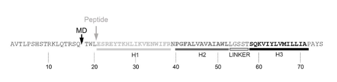
To focus the study on the functionally relevant region of the protein, a 55-residue peptide fragment was designed corresponding to residues 21 to 75 of the mature M protein, omitting the disordered N-terminal loop. The retained sequence encompassed all three helical regions of M, including the two transmembrane domains that naturally embed within the virion membrane. This truncated yet structurally representative fragment served as a well-defined model for investigating the membrane interactions of M, oligomeric assembly and potential pore-forming activity. The peptide sequence was derived from a clinically relevant New World ZIKV isolate (PE243), isolated during the 2015 Brazilian epidemic.
Purifying Highly Hydrophobic Peptides
The M peptide sequence presented significant synthesis and purification challenges due to its high hydrophobicity. Two sequence motifs were particularly problematic:
- VAVAIA: A stretch rich in valine, alanine, and isoleucine residues with bulky nonpolar side chains that repel water and promote intermolecular aggregation
- WIFR: A section containing tryptophan and phenylalanine, further increasing hydrophobicity and aggregation propensity
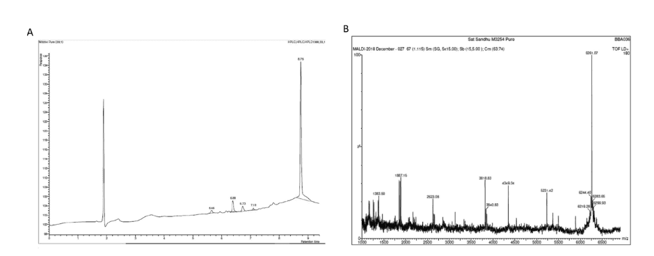
Together, these motifs can lead to premature truncation during synthesis, poor solubility of the crude product, and difficulty achieving adequate resolution during standard reversed-phase purification. To address this, the crude peptide was purified using dialysis rather than conventional chromatographic approaches. This strategy selectively removed small molecular weight contaminants and synthesis by-products while retaining the full-length peptide. As a result, the final product achieved >95% purity, as confirmed by HPLC and mass spectrometry, which was sufficient for all downstream studies.
Why Acetylate the N-Terminus?
The M peptide was synthesised with an acetylated N-terminus to improve the physiological relevance of the experimental system. In its native form, M is embedded within the lipid bilayer as part of a continuous polypeptide. A synthetic peptide naturally carries a free amine at its N-terminus, introducing an artificial charge absent in the native protein fragment. Peptide acetylation neutralises this charge, causing the peptide to more closely mimic an internal membrane-embedded protein segment.
Therefore, the benefits of N-terminal peptide acetylation include:
- Reduced non-physiological electrostatic interactions at membrane surfaces
- Increased the stability of the α-helical structure in lipid environments
- Improved peptide insertion and membrane orientation
- Enhanced physiological relevance of channel formation experiments
Results: ZIKV M Protein Forms Functional Ion Channels That Can Be Drugged
Synthetic Peptides M Exhibit Viroporin-Like Assembly
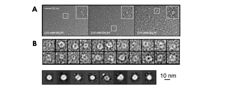
First, the group solubilised synthetic M peptides in DH6PC, a detergent that forms monolayered micelles and prevents non-specific aggregation. Negative-stain electron microscopy revealed that at lower detergent concentrations, the synthetic peptide M assembled into discrete, circular, channel-like structures, a key morphological hallmark of viroporins. While some structural variability was observed, the formations were clearly non-aggregated and consistent with membrane-spanning oligomeric complexes.
Observation of Higher-Order Oligomers on Native PAGE
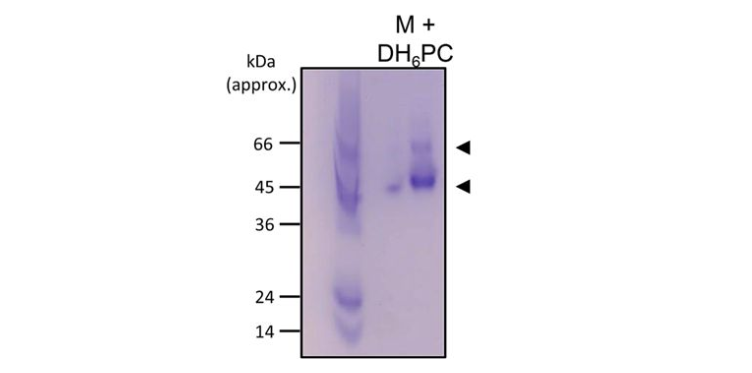
Native polyacrylamide gel electrophoresis (PAGE) in 300 mM DH6PC provided further evidence of oligomerisation. The dominant species migrated at approximately 45–50 kDa, with a minor band near 60 kDa. These molecular weights were consistent with hexameric or heptameric assemblies and confirmed the possible formation of higher-order oligomers well beyond simple dimers, a defining characteristic of functional viroporins.
Ion Channel Activity Confirmed in Liposomes
To test whether these oligomers were functionally active as ion channels, the team employed a dye-release liposome assay. M peptides triggered dose-dependent release of encapsulated fluorescent dye at sub-micromolar concentrations, indirectly confirming channel activity.
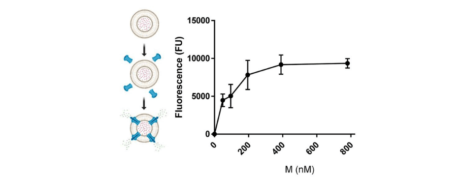
Notably, slightly acidic conditions designed to mimic the pH of the endosomal lumen, significantly enhanced dye release. This pH-dependent activation mirrors the behaviour of well-characterised viral ion channels, including influenza A M2, hepatitis C p7, and HPV E5, and suggests that M channel activity may be physiologically triggered during endosomal viral entry.

Activity Preserved in Highly Curved Membranes
Viroporins must function within the tightly curved membranes of viral envelopes. To assess this, the synthetic peptides were tested in small, highly curved liposomes that closely approximate envelope geometry. Channel activity was retained under these conditions, though dye release was marginally slower. This was likely attributable to a higher liposome-to-peptide ratio rather than any loss of function. This demonstrated that M channel formation was possible across physiologically relevant membrane geometries.
Rimantadine Blocked Channel Activity
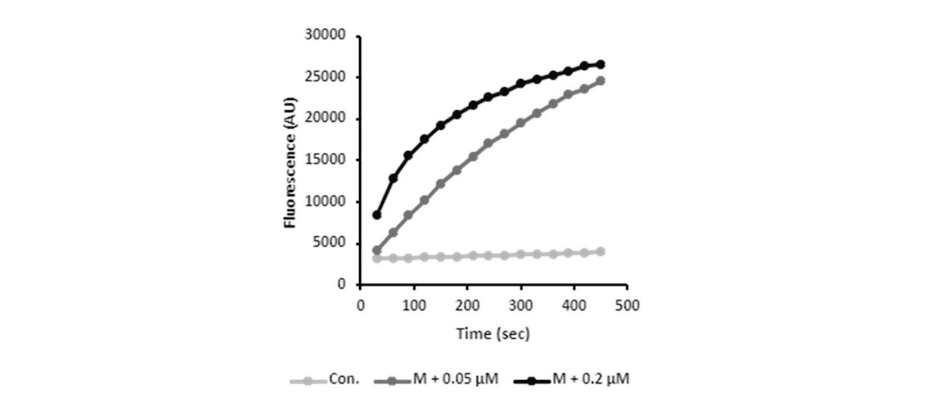
Finally, the researchers tested whether peptide M could be targeted pharmacologically with an inhibitor known to block influenza M2 viroporins. Treatment with 1 µM rimantadine substantially reduced M-mediated dye release. This indicated that the M peptide could fold into a stable channel structure that could be drugged similarly to other viral targets.

Rapid Throughput Small Molecule Screening Using a Peptide-Based Assay
Building on these findings, molecular dynamics simulations were used to generate structural models of the M channel and identify potential druggable binding sites. These models then served as the basis for in silico screening of repurposed drugs and bioactive compounds. The selected hits were subsequently evaluated using the peptide-based channel assay. This led to the identification of several inhibitors with superior qualities to rimantadine, which were then successfully tested in a cell-based plaque assay.
Conclusion
This case study illustrates how synthetic peptides can provide key insights into the biology of challenging membrane proteins and accelerate early-stage drug discovery. The synthetic M peptides enabled key mechanistic insights that would have been difficult to achieve with the full-length protein. With them, the group confirmed that the M protein could self-assemble into functional ion channels, respond to acidic pH and be inhibited by small molecules. These findings directly supported the identification of M as a drug target and laid the groundwork for subsequent antiviral studies in cell culture and preclinical models. The full study can be read here:
Inhibitors of the small membrane (M) protein viroporin prevent Zika virus infection | eLife.
“We were delighted to support this research. Projects like this, where synthetic peptides are used to better understand the function of a challenging membrane protein and drive early antiviral drug discovery, are exactly the kind of work we are passionate about at AltaBioscience. Delivering a high-purity hydrophobic peptide that performed reliably across such a diverse range of biophysical and functional assays was a rewarding challenge, and we look forward to supporting the next stages of this important research.”
Sat Sandhu, Head of Peptide Chemistry at AltaBioscience.
At AltaBioscience, we specialise in delivering high-purity, custom-synthesised peptides tailored to your research needs. We have extensive experience in complex and challenging sequences, including long hydrophobic peptides that are difficult to synthesise and purify by conventional methods. Our capabilities span a wide range of chemical modifications and conjugations, allowing us to design peptides that closely mimic native protein’s structural motifs. Whether you are investigating membrane protein function, validating a novel drug target or developing inhibitor screening assays, we have the expertise to support all your projects.
Contact us to learn more.