Epitope Mapping: Methods, Applications and Peptide-Based Approaches
Jump to section
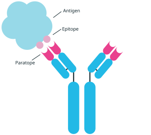
Epitope mapping is the process of identifying the specific region of an antigen, known as the epitope, that an antibody recognises and binds. Knowing where an antibody binds provides the molecular detail needed to optimise and further develop it, protect it commercially, and predict how it will behave.
That detail is critical for antibody and vaccine research. Two antibodies raised against the same target can bind different epitopes and differ widely in function1. As such, mapping the binding site is invaluable for guiding lead selection, identifying cross-reactivity, and increasingly underpins the patent and regulatory data packages behind therapeutic programmes2.
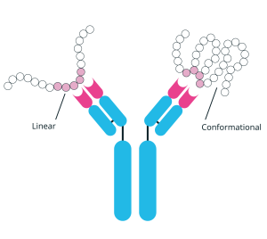
Epitopes are either linear, consisting of a continuous stretch of amino acids, or conformational and so formed by residues brought together when the protein folds. Linear epitopes are the most tractable to map, and this is where peptide methods excel: overlapping peptides tiled across a sequence can be screened on a peptide microarray or tested as peptide pools, at scale. Conformational epitopes are resolvable only by structural methods such as X-ray crystallography or cryo-EM.
This guide covers how linear epitope mapping works, the peptide tools behind it, its key applications and the practical factors that keep the resulting data reliable.
How Linear Epitope Mapping Works
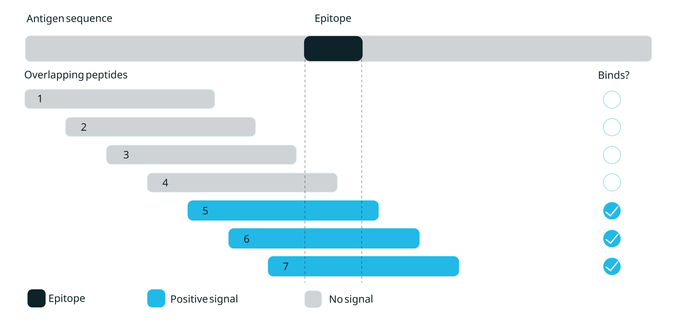
Linear epitope mapping works on a simple principle: break a target protein into short, overlapping peptide fragments, present them to an antibody or immune cell, and see which fragments are recognised. A positive signal means the epitope (or part of it) lies within that peptide, so the binding site can be located along the sequence without needing the intact, folded protein.
Library design is one of the most important factors in the success of a linear epitope mapping study. It shapes both the resolution of the map and the reliability of the data. Peptide length, the overlap between neighbouring peptides, and how the library is read out are all important factors to consider.
Overlapping Peptide Libraries
The foundation is a set of short peptides (typically 10–20 amino acids) that together cover the whole protein sequence. This overlapping scan is normally the first pass, locating the region an antibody or T cell recognises before finer methods pinpoint the exact residues.
Each peptide starts a fixed number of residues further along than the last, a step known as the offset, so that neighbouring peptides overlap and no part of the sequence falls in a gap between them. A smaller offset gives finer resolution but requires more peptides3.
Peptide length is matched to what’s being mapped. T-cell epitopes are short and tightly sized, so T-cell libraries use correspondingly short peptides. Antibody (B-cell) epitopes vary more, and are therefore screened across a range of lengths.
Alanine and Substitution Scanning
Once a peptide from the overlapping screen scores positive, substitution scanning identifies the binding residues within it. Each residue in the peptide is replaced in turn, usually with alanine, which removes the residue’s side chain while leaving the backbone intact. Next, the variants are re-tested.
A drop in binding marks that residue as part of the epitope, and in this way, it is possible to map the residues that make up the epitope. Since the variants are chemically synthesised, rather than made by mutating and expressing the whole protein, every sequence is defined exactly and no cloning step is needed4.
Pooling and Deconvolution
For large libraries or functional T-cell assays, peptides are tested in pools rather than one by one, and positive pools are then deconvoluted to find the active peptide. Matrix pooling can cut the number of assays several-fold without losing sensitivity5.
Each of these steps builds on the last: overlapping libraries locate the reactive region, microarrays screen it at scale, substitution scanning narrows it to individual residues, and pooling keeps large or functional assays manageable. Conformational epitopes, which peptides cannot reproduce, require alternative structural methods.
Applications of Epitope Mapping
Epitope mapping is valuable wherever knowing the exact binding site informs a decision: which antibody to take forward, whether a vaccine target will work, or how to tell one pathogen apart from another.
Antibody epitope mapping
For therapeutic and diagnostic antibodies, the binding site is part of the product. Antibody epitope mapping shows where on the antigen an antibody binds, which supports several decisions at once: differentiating lead candidates, checking cross-reactivity with related proteins, and building the binding-site evidence that increasingly underpins antibody patents6.
Application: From Polyclonal Serum to Monospecific Antibodies.
Epitope mapping can also be a manufacturing step. In one published workflow, a polyclonal antiserum was screened against a peptide array to locate its linear epitopes; those peptides were then coupled to columns and used to affinity-purify a panel of monospecific antibodies, each specific to a different epitope 1.
Mapping the epitopes first enabled the rational selection of the most specific, best-performing reagents for downstream applications, removing the need to generate and screen large antibody panels by trial and error. This is both faster and more cost-effective. The same approach could also revalidate and optimise existing polyclonal antibody reagents, converting them into better-defined, monospecific ones.
For researchers wanting to follow this protocol to generate monospecific antibodies from a polyclonal antiserum, we can synthesise peptide libraries and peptide-antigen affinity columns once the peptide epitopes have been identified. For more information, please view our custom peptide services.
Vaccines and immunotherapy
In vaccine and immunotherapy pipelines, epitope mapping identifies the peptides that drive a protective T-cell response. That supports epitope-based vaccine design, the validation of tumour neoantigens for personalised cancer vaccines, and immune monitoring: tracking a patient’s antigen-specific T-cell responses across the course of treatment7.
Infectious diseases
Mapping the epitopes a pathogen exposes to the immune system informs both vaccine targets and diagnostics. Novel CD8+ T-cell epitopes have been mapped in viruses such as human herpesvirus 6B to help guide T-cell therapies for transplant patients8.
For diagnostics, synthetic peptides offer a purer, more standardised alternative to crude pathogen extracts. For example, a screen for Echinococcus infection using a peptide microarray supplied by AltaBioscience found that pooling several peptide antigens improved sensitivity over any single peptide9.
Practical Considerations For Epitope Mapping
Whether a linear epitope mapping result holds up depends on the peptides behind it and the format in which they are used. These practical factors are worth careful consideration and consultation with a peptide supplier:
Choosing an Epitope Mapping Strategy
There are several questions to consider before settling on a design:
- What is being mapped? An antibody’s binding site (a B-cell epitope) and a T-cell response tend to call for different libraries and readouts: binding assays, which detect whether an antibody attaches to a peptide, for the former; functional cell-based assays, which measure a T-cell response, for the latter.
- What resolution is needed? An overlapping scan locates the reactive region; adding a substitution scan narrows it to the individual binding residues. Region-level is often enough to tell antibodies apart, while residue-level is needed for fine characterisation or IP evidence.
- Is the epitope likely linear? If an antibody loses binding to a denatured antigen, or a full linear scan comes back negative, the epitope may well be conformational; in which case a structural method is usually the better route.
Choosing the Best Peptide Format
Peptides can be presented in two main ways, and the choice follows the biology of the assay rather than a fixed rule:
- Peptide microarrays immobilise the library on a slide and screen thousands of sequences in parallel from a single sample; well suited to high-throughput antibody mapping across whole proteins or panels.
- Solution-phase pools in microplates suit functional assays such as T-cell readouts by ELISpot, where the peptides need to reach live cells.
AltaBioscience’s EpiScan technology generates the overlapping peptides for either route, supplied as libraries, pools or microarray slides. Learn more about EpiScan.
Matching Purity to the Assay
Synthesis by-products — truncated or deletion sequences, and residual protecting groups — can bind non-specifically and generate false positives. How much this matters depends on the assay: higher purity means fewer artefacts, but it also means more purification, higher cost per peptide and longer turnaround, which quickly adds up across a library of hundreds. Purity should therefore be matched to what the assay demands rather than maximised by default:
- Discovery screening: crude or desalted peptides (often >70%) are enough to find hits cheaply across large libraries.
- Binding assays: at least 85% purity is preferred for reliable ELISA or array data.
- Functional and quantitative assays: purified, fully characterised peptides (>90–95%) are needed for immune monitoring and any clinical use10.
Advantages and Limitations of Linear Epitope Mapping
Both linear peptide mapping and structural methods address the same underlying question: where does an antibody bind? But their approaches and outputs differ. Linear peptide mapping can identify continuous (linear) epitopes, while structural methods are able to resolve conformational epitopes that depend on the three-dimensional arrangement of residues. They each have advantages and limitations. Most epitope work draws on both.
Peptide-based mapping | Structural methods | |
Epitope type | Linear only | Conformational and linear |
Methods | Overlapping libraries, microarrays, substitution scans | X-ray crystallography, cryo-EM, HDX-MS |
Throughput | High — thousands of peptides in parallel | Low — one antibody–antigen complex at a time |
Resolution | Residue-level along the sequence | Full three-dimensional detail |
Sample | Works on denatured or folded targets | Needs an intact, folded antigen (or complex) |
Cost and speed | Lower cost, faster | Higher cost, slower |
Best for | Antibody screening, T-cell epitopes, diagnostics | Defining discontinuous binding sites |
Linear epitope mapping is a fast, scalable way to pin down exactly where antibodies and T-cells recognise their targets.
AltaBioscience provides custom overlapping libraries, peptide pools and microarrays for linear epitope mapping through its Peptide Libraries and Microarrays service, each synthesised and quality-controlled by HPLC and mass spectrometry in an ISO 9001-certified UK laboratory.
Contact our team to talk through your target and scope the right mapping approach.
References
- Hjelm, B. et al. Generation of monospecific antibodies based on affinity capture of polyclonal antibodies. Protein Science 20, 1824–1835 (2011).
- Deng, X., Storz, U. & Doranz, B. J. Enhancing antibody patent protection using epitope mapping information. MAbs 10, 204–209 (2018).
- Forsström, B. et al. Proteome-wide epitope mapping of antibodies using ultra-dense peptide arrays. Molecular and Cellular Proteomics 13, 1585–1597 (2014).
- Buus, S. et al. High-resolution mapping of linear antibody epitopes using ultrahigh-density peptide microarrays. Molecular and Cellular Proteomics 11, 1790–1800 (2012).
- Fiore-Gartland, A. et al. Pooled-Peptide Epitope Mapping Strategies Are Efficient and Highly Sensitive: An Evaluation of Methods for Identifying Human T Cell Epitope Specificities in Large-Scale HIV Vaccine Efficacy Trials. PLoS One 11, e0147812 (2016).
- Deng, X., Storz, U. & Doranz, B. J. Enhancing antibody patent protection using epitope mapping information. MAbs 10, 204–209 (2018).
- Sonntag, K. et al. Immune monitoring and TCR sequencing of CD4 T cells in a long term responsive patient with metastasized pancreatic ductal carcinoma treated with individualized, neoepitope-derived multipeptide vaccines: a case report. Journal of Translational Medicine 2018 16:1 16, 23- (2018).
- Halawi, M., Khan, N. & Blake, N. Identification of novel cd8+ t cell epitopes in human herpesvirus 6b u11 and u90. Immun. Inflamm. Dis. 3, 118–131 (2015).
- List, C. et al. Serodiagnosis of Echinococcus spp. Infection: Explorative Selection of Diagnostic Antigens by Peptide Microarray. PLoS Negl. Trop. Dis. 4, e771 (2010).
- Castro, A. et al. Peptide pools for target antigen identification, immune monitoring, and cellular therapy. Cytotherapy 22, S119–S120 (2020).