Fluorescent Peptides: A Guide for Life Science Researchers
Fluorescent peptides have become invaluable tools in life sciences, enabling researchers to track molecular interactions, visualise cellular processes, and develop innovative assays. Whether you are designing fluorescence resonance energy transfer (FRET) peptides or simply labelling peptides for imaging studies, understanding the key factors influencing their performance is crucial.
In this latest article, we explore common fluorescent dyes, important design considerations, techniques using peptide-based fluorescence probes, and best practices for dissolving, handling and storing them. Read more below or download our complete fluorescent peptide guide.
For any custom peptide synthesis enquiries, please email info@altabioscience.com.
Which Dyes Are Used for Fluorescent Peptide Labelling
Fluorescent peptides are synthetic peptides conjugated to fluorescent dyes. These dyes are organic molecules with a conjugated π-electron system that allows them to absorb and emit light at specific wavelengths. They typically contain benzene and heterocyclic rings, with their structures determining their spectral properties, brightness, and stability. Therefore, choosing the right fluorescent dye is essential for optimising experimental outcomes. Here are some of the most commonly used dyes in peptide-based assays:
- Carboxyfluorescein (FAM): It is a popular green fluorophore (excitation/emission: ~494/518 nm) used in various biological assays. This is by far the most commonly used and cost-effective dye. Its absorption wavelength is a very close match to the 488nm emission line of an argon ion laser. It can be easily incorporated into both peptides and oligos at virtually any position with few chemical conflicts. The primary drawbacks are the tendency of fluorescein to photo bleach and to have a relatively narrow working range of pH 7.5-8.5.
- Fluorescein isothiocyanate (FITC) is another fluorescein derivatives that share similar spectral properties as FAM, excitation around 495 nm and emission near 517–519 nm. Its isothiocyanate group reacts readily with primary amines to form stable thiourea bonds, making it suitable for labelling proteins, antibodies, and peptides.
- Edans (5-((2-Aminoethyl)amino)naphthalene 1-sulfonic acid) max Abs = 335nm, max Em = 493nm. It is a well-used donor molecule for FRET based assays which is usually paired with Dabcyl. Dabcyl however, is not fluorescent due to its chemical structure, but has been designed to absorb light at a specific wavelength but not reemit it as fluorescence. Instead, Dabcyl dissipates this energy as heat rather than reemitting it as light, hence “quenching” the donor’s fluorescence. As a result, this interaction enables researchers to monitor molecular interactions or conformational changes based on the changes in fluorescence intensity.
- Tetramethylrhodamine (TAMRA): A red fluorophore (excitation/emission: ~557/583 nm) often used in FRET studies. Tamra is readily added to peptides and can be positioned anywhere along the peptide chain. Tamra gives a high intensity fluorescent response, is reasonably photo stable and has a wide pH range. In addition, its intense purple colour makes for easy visual location of the compound.
- DyLightTM dyes: These are a range of 9 high performance dyes developed by Pierce Biotechnologies, covering a wide absorption and emission range. Advantages include narrow spectra which allows the use of dye multiplexing, good photo stability, wide working pH range and intense fluorescence. The DyLight dyes cover an excitation range of 400nm to 770nm with emission over 420nm to 778nm.
- Cyanine Dyes (Cy3, Cy5, Cy7): These dyes are extensively utilised for labelling peptides, proteins, and nucleic acids. They offer a range of spectral properties for multiplexing applications. For instance, Cy3 has excitation/emission maxima at approximately 550/570 nm, Cy5 at 650/670 nm, and Cy7 at 745/800 nm.
- Alexa Fluor Dyes are a series of highly photostable and bright fluorescent dyes, spanning the visible to near-infrared spectrum. They are water-soluble and maintain fluorescence across a broad pH range, making them ideal for labelling peptides in various biological applications.
- BODIPY Dyes are known for their sharp emission peaks, high quantum yields, and environmental insensitivity, however, unstable to TFA. They are suitable for lipid and membrane studies due to their hydrophobic nature.
The choice of fluorescent dye can significantly impact the performance and outcome of your experiment. With a variety of dyes available, each offering different advantages, it is important to select the one that best fits your specific research requirements.
Download our comprehensive Fluorescent Peptide Guide, featuring a detailed comparison table of popular fluorescent dyes used in peptide labeling. Explore key properties including excitation and emission wavelengths, brightness, photostability, pH sensitivity, and cost.
7 Key Considerations When Designing Fluorescent Peptides
When incorporating fluorescent labels into peptides, we recommend considering the following factors to ensure optimal performance:
1. Fluorophore Selection
Selecting the appropriate fluorophore depends on the optical properties of your imaging or detection system and the following factors:
- Desired excitation and emission wavelengths, which refers to the wavelengths at which a fluorophore absorbs and emits light, respectively.
- Photostability, e.g. how resistant a fluorophore is to photobleaching, which is the irreversible loss of fluorescence due to prolonged light exposure. In particular, high photostability ensures longer-lasting signals, making it essential for imaging applications.
- Brightness is a measure of how intense the emitted fluorescence is, which depends on both the fluorophore’s quantum yield (efficiency of fluorescence) and its molar extinction coefficient (ability to absorb light).
- and quenching susceptibility.
Consider the optical properties of your imaging or detection system when making a choice.
2. Spacer Inclusion
In many cases, a spacer between the peptide and the dye can reduce steric hindrance, minimise potential interference with biological activity or to avoid unwanted side reactions with the peptide. For instance, introducing FITC at the N-terminus of a peptide when on solid phase support results in the truncation of the terminal amino acid during the acidic cleavage (Jullian, 2009).
This well-studied side reaction can be avoided by either coupling the dye with a lysine or ornithine side chain after selective deprotection, or when performing the reaction by solid-phase peptide synthesis by introducing a spacer such as:
- Aminohexanoic acid (Ahx), a flexible alkyl six-carbon linker.
- β-Alanine, a minimal spacer that maintains peptide conformation.
Peptide PEGylation with polyethylene glycol (PEG) is another example of an inert spacer known to enhance solubility due to its highly hydrophilic nature.
3. Dye Positioning: Where Should You Label the Peptide?
Peptide conjugation to fluorescent probes is most commonly achieved through amine chemistry or by targeting cysteine residues, allowing for labelling at the N-terminus or on the side chains of lysine or cysteine.
A key consideration when designing fluorescent peptides is deciding where to position the dye to maintain the desired biological activity and structural integrity.
- N-terminal labelling: Selective N-terminal labelling can be challenging in the presence of lysine residues, which also contain reactive amino groups. Strategies such as temporary side-chain protection, adjusting the pH to favour N-terminal reactivity (pKa ~8) instead of lysine’s ε-amino group (pKa ~10.5), or the use of selective reagents can help ensure dye attachment specifically at the N-terminus (Chen, 2017).
- If labelling of all available amino groups is required, non-selective amine-reactive dyes can be used to modify both the N-terminus and lysine side chains, resulting in multiple conjugation sites.
- To preserve a free N-terminus and label the C-terminus instead, alternative chemistries such as coupling to a C-terminally introduced cysteine or modifying a terminal carboxyl group (with appropriate activation) can be employed.
Careful selection of the labelling strategy is essential to ensure site-specificity, maintain peptide function, and achieve consistent results in downstream applications.
Case Study: Site-Specific FITC Labelling of Histone H4 (1–21) Peptide
Peptide Sequence: SGRG-K(Ac)-GG-K(Ac)-GLGKGGA-K(Ac)-RHRKV-K(ahx-FITC)-amide
Challenges: Synthesis of a fluorescently-labelled histone peptide containing:
- One FITC-labeled lysine at the C-terminus via an Ahx spacer
- in the presence of three internal acetylated lysines (K(Ac)).
Purity required >95%, 5mg
Strategy:
- We used Fmoc-Lys(Ac)-OH for site-specific incorporation of acetylated lysines.
- We introduced Dde-protected Lys at C-terminus to enable orthogonal deprotection and selective FITC labeling.
- We employed Boc-Ser at N-terminus to avoid interference with Dde deprotection.
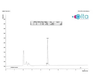
Results:
The target peptide was successfully synthesised with a final purity of 98.8%, as confirmed by HPLC (Fig.1). The identity of the peptide was also verified by mass spectrometry (Fig. 2), confirming the expected molecular weight.

Note: MALDI-ToF analysis led to partial fragmentation of the FITC group during ionisation. However, the FITC-Peptide was confirmed using Electrospray mass spectrometry, where the single target molecule was observed.
4. How to Prevent Self-Quenching and Aggregation
High concentrations of fluorescent dyes or closely spaced fluorophores can lead to self-quenching and aggregation, resulting in a significant reduction in fluorescence intensity. This effect is particularly relevant in fluorescence microscopy applications, such as confocal laser scanning microscopy (CLSM), which is commonly used to examine the subcellular localisation of fluorescent biomolecules such as cell-penetrating peptides labelled with fluorescein or rhodamine B.
In such experiments, an excessive accumulation of fluorescent peptides within living cells may lead to diminished or absent signal, potentially causing misinterpretation of the data as a lack of target presence rather than quenching-related signal loss. However, this can be prevented by diluting the fluorescent peptide with a non-fluorescent (e.g. acetylated) analogue. This reduces local fluorescent peptide concentrations, dramatically lowering self-quenching without altering peptide function (Swiecicki, 2016).
5. Impact of the Dyes on the Fluorescent Peptide’s Physicochemical Properties
Fluorescent dyes are often inherently hydrophobic, which can significantly influence the physicochemical properties of the labelled peptides. Incorporating these dyes may alter the peptide’s conformation, flexibility, and overall charge distribution, potentially affecting its interaction with the protein targets, lipid membranes, cellular uptake, and subcellular localisation (Bechara, 2013). Several studies have shown that such modifications can impact biological activity and experimental outcomes. As a result, these factors should be carefully considered during fluorescent peptide design and experimental planning to ensure accurate data interpretation and reliable results.
6. Choosing the Right Dye Isomer
Fluorescent dyes such as fluorescein and Tamra exist as two isomers. For most purposes, where the dye is simply being used as a tracer, the chemically identical, mixed isomers can be used. This greatly reduces the overall cost of the synthesis. Only where spectral studies are being performed is it important to consider using the discrete isomers.
7. pH Sensitivity of Fluorescent Dyes
The fluorescence intensity of certain dyes can be significantly influenced by pH, which is crucial when designing fluorescent peptides for environments with variable acidity. For instance, dyes like FITC and FAM exhibit decreased fluorescence in acidic conditions, potentially leading to underestimation of peptide presence or activity in compartments such as endosomes or lysosomes. This pH-dependent variability can compromise data accuracy, especially in live-cell imaging or intracellular tracking. To mitigate such issues, we recommend selecting pH-stable dyes like Alexa Fluor 488 or DyLight 488, as they maintain consistent fluorescence across a broad pH range, ensuring reliable signal detection regardless of the cellular environment.
How to Label Your Peptide with a Fluorescent Dye
If you’ve already synthesised a peptide and now wish to label it with a fluorescent dye, there are several approaches to consider, depending on the peptide’s current modifications and your experimental needs.
Utilising Biotinylated Peptides with Fluorescent Streptavidin
If your peptide is already biotinylated, an efficient method to introduce fluorescence is by binding it with a fluorescently labelled streptavidin. The biotin-streptavidin interaction is one of the strongest non-covalent bonds, ensuring stable complex formation. One advantage of this method is that various fluorophores can be used without modifying the peptide directly. However, the addition of streptavidin increases the molecular weight, which might affect the peptide’s behaviour in certain assays
Direct Chemical Labelling of Peptides
If your peptide lacks biotin or you prefer a smaller label, we can perform direct chemical conjugation of a fluorophore to the peptide as an alternative method.
Common methods include:
- NHS Ester Chemistry: Targets primary amines (e.g., lysine residues or N-terminus) to form stable amide bonds with the fluorophore.
- Maleimide Chemistry: Reacts with thiol groups (e.g., cysteine residues) to form thioether bonds.
However, while post-synthetic labelling via biotin-streptavidin systems or by direct chemical conjugation is effective and flexible, it can introduce variability, reduce yield, or require additional purification steps. In many cases, we recommend incorporating the fluorophore during peptide synthesis as this is the most efficient and reliable approach, ensuring precise labelling, consistency, and time savings.
Examples of Fluorescence-Based Techniques Using Fluorescent Peptides
Fluorescent peptides are highly versatile tools, valued for their excellent biocompatibility, specificity, and low toxicity. They are widely used in imaging, disease detection, ion sensing, and biomolecular analysis for real-time monitoring and multiplexing (Wang, 2023). In particular, fluorescent probes enable precise investigation of molecular interactions, tracking of subcellular processes, and quantification of biological responses with exceptional sensitivity and specificity. Below are some of the most widely used techniques that leverage fluorescent peptides:
- Fluorescence Microscopy (CLSM, STED)
Confocal Laser Scanning Microscopy (CLSM) and super-resolution methods like Stimulated Emission Depletion (STED) microscopy are essential for visualising the intracellular distribution of fluorescent peptides. CLSM offers excellent optical sectioning to examine peptide localisation in specific organelles or cellular compartments, while STED allows for imaging at the nanometre scale, surpassing the diffraction limit of light.
- Fluorescence Resonance Energy Transfer (FRET)
FRET-based assays are widely used to probe protein–peptide interactions and conformational changes. By labelling peptides with a donor–acceptor fluorophore pair, researchers can detect changes in proximity or binding events based on shifts in emission intensity. This is ideal for studying dynamic molecular interactions in real-time.
- Flow Cytometry
Fluorescently labelled peptides can be quantified in large cell populations using flow cytometry. This technique is particularly useful for assessing cell surface binding, cellular uptake (Kroemer, 2002), or receptor-ligand interactions, and allows for high-throughput analysis of single cells based on fluorescence intensity.
- Fluorescence Polarisation (FP) Assays
FP assays utilise fluorescent peptides to measure binding affinity between a small labelled molecule and a larger binding partner. Upon binding, the slower rotation of the complex results in higher polarisation of emitted light, providing a reliable and quantitative readout of molecular interaction strength.
- Live Cell Imaging
Live cell imaging enables the real-time visualisation of fluorescent peptides inside living cells, offering insights into cellular uptake, trafficking, and subcellular localisation. This is especially valuable in drug delivery research and studies on intracellular signalling pathways.
- High-Content Screening (HCS)
In drug discovery and systems biology, HCS integrates fluorescence imaging with automated data analysis to evaluate large libraries of compounds or peptides. Fluorescent peptides are commonly used to assess phenotypic changes, localisation patterns, or signal pathway activation in response to treatment.
- NIR-fluorescence Imaging
It is a powerful, non-invasive technique used in biomedical research for real-time tracking of biological processes in vivo (Xu, 2022). It offers deep tissue penetration, low background interference, and high sensitivity. Fluorescent probes labelled with NIR dyes are particularly useful as they combine the targeting specificity of peptides with the imaging capabilities of NIR fluorophores, enabling precise visualisation of cellular and molecular targets, such as tumours or inflamed tissues.
Handling and Storage of Fluorescent Peptides
Proper handling and storage are critical for maintaining the stability and fluorescence intensity of labelled peptides:
- Storage Conditions: Store fluorescent peptides at -20°C or lower, protected from light and moisture.
- Solvent Considerations: Many fluorophores are sensitive to oxidation; use anhydrous solvents when preparing stock solutions.
- Aliquoting: Divide peptides into small aliquots to minimise freeze-thaw cycles and degradation.
- Protection from Light: Use amber vials or foil-wrapped tubes to prevent photobleaching.
- Dissolution of fluorescent peptides: The solubility of peptides is highly dependent on their sequence. The addition of fluorescent dyes often increases the hydrophobicity of peptides, potentially complicating their dissolution. To enhance solubility, especially for hydrophobic peptides, we employ co-solvents such as dimethyl sulfoxide (DMSO) or acetonitrile. In addition, peptides that are acidic, i.e. contain more acidic amino acids than basic, will be more soluble at higher pH and vice versa, peptides that are overall basic will be most soluble at lower pH.
By incorporating these best practices and considerations, you can maximise the effectiveness of fluorescent peptides in your research.
Custom Fluorescent Peptide Synthesis
At AltaBioscience, we specialise in custom peptide synthesis, including fluorescently labelled peptides tailored to your research needs. Our experts can assist in selecting the right fluorophore, spacer, and labelling strategy for your specific application.
Should you require further information, contact us at info@altabioscience.com for expert support.
Download our Fluorescent Peptides Guide

References
Bechara, C. and Sagan, S., 2013. Cell-penetrating peptides: 20 years later, where do we stand? FEBS Letters, 587(12), pp.1693–1702. https://doi.org/10.1016/j.febslet.2013.04.031
Chen, D., Disotuar, M.M., Xiong, X., Wang, Y. and Chou, D.H.-C., 2017. Selective N-terminal functionalization of native peptides and proteins. Chemical Science, 8(4), pp.2717–2722. https://doi.org/10.1039/c6sc04744k.
Jullian, M., Hernandez, A., Maurras, A., Puget, K., Amblard, M., Martinez, J. and Subra, G., 2009. N-terminus FITC labeling of peptides on solid support: the truth behind the spacer. Tetrahedron Letters, 50(3), pp.260–263. https://doi.org/10.1016/j.tetlet.2008.10.141.
Kroemer, G., 2002. The role of the mitochondrial permeability transition pore in cell death. Biochimica et Biophysica Acta (BBA) – Bioenergetics, 1555(1–3), pp.73–76. https://doi.org/10.1016/S0005-2736(02)00471-6.
Swiecicki, JM., Thiebaut, F., Di Pisa, M. et al. How to unveil self-quenched fluorophores and subsequently map the subcellular distribution of exogenous peptides. Sci Rep 6, 20237 (2016). https://doi.org/10.1038/srep20237.
Wang, B., Wang, L., Chen, C., Zhang, Y., Gao, J., Lu, K., Yan, C., Nan, G. and Li, Y., 2023. A review on recent advances in peptide-based fluorescence probes and their potential applications. ChemistrySelect, [online] Available at: https://doi.org/10.1002/slct.202302216
Xu, H., Wang, H., Xu, Z., Bian, S., Xu, Z. and Zhang, H., 2022. The multifaceted roles of peptides in “always-on” near-infrared fluorescent probes for tumor imaging. Bioorganic Chemistry, 129, 106182. https://doi.org/10.1016/j.bioorg.2022.106182


